The impact of repetitive head injuries in contact sports and development of Chronic Traumatic Encephalopathy (CTE)
For many decades it has been recognised that contact sports such as boxing can lead to chronic progressive brain damage and neurological symptoms, even progressing to dementia.
Chronic Traumatic Encephalopathy (CTE), is a progressive neurodegenerative disease caused by repetitive blows to the head and concussions. CTE can lead to dementia and related physical and cognitive symptoms such as memory problems, a decline in thinking ability, confusion, aggression, depression and changes in personality; all of which can be debilitating and life-changing for those affected.
The first case of repetitive head injury associated with brain damage was diagnosed in a retired NFL player 15 years ago and the term Chronic Traumatic Encephalopathy (CTE) was introduced.
Over the past 15 years there has been an explosion of interest in the potential role and long-term consequences of repetitive head injury, in contact sports.
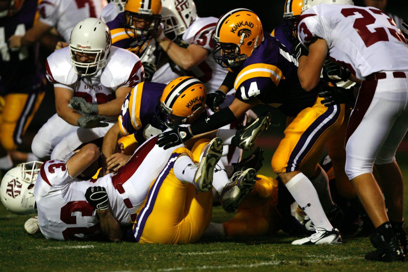
Much of the work done on CTE has been focused on American football players and in 2017 alarming statistics were published which included:
Former NFL players aged 30–49 demonstrated memory-related diseases, 19 times more frequently than individuals, in the same age group, in the general population.
6.1% of NFL players over the age of 50 were diagnosed with dementia , which is 5x the national average of 1.2%.
In 2016, the NFL acknowledged CTE as a disease linked to repeated brain trauma and associated memory loss, depression and progressive dementia.( Mar. 15, 2016 10:58 AM EDT
http://bigstory.ap.org/urn:publicid:ap.org:7fd994cea9734899a27ef57ffc45c7e6)
Furthermore in 2017, an NFL official formally acknowledged a link between (American) football and CTE, for the first time.
50 years after England’s World Cup Win – 50% of England’s surviving football players were suffering from dementia or memory loss.
With an increased interest in this condition, further cases of CTE have been diagnosed, clinically, in soccer, rugby union, rugby league and Australian rules football. It’s understandable why players and sports governing bodies are increasingly worried about this hugely concerning issue.
In addition, large epidemiological studies have shown the risk of dementia and other neurodegenerative diseases such as Motor Neurone Disease are four times higher in players of contact sports.
There is a very variable incidence of concussion and sub concussion caused by contact sport; Rugby Union has the highest incidence, soccer is at a much lower level. This is likely to be important because in the largest post mortem studies of NFL players there appears to be a clear dose-response curve, the lower you play and higher level at which you play, the higher the chance of developing CTE.
At the moment, despite a large wave of coherent research pointing directly to the risk of developing CTE from contact sports, controversy remains. One side of the argument says that it’s too early to conclude that there is a link, the other says that there is enough evidence to act now.
At Re:Cognition Health our perspective is that high quality clinical and diagnostic assessments can unlock this debate. In our experience, it is possible to identify high risk individuals and form a combination of assessments to offer a reasonably certain clinical probability of a likely diagnosis. It is also crucial that any players who are struggling, regardless of the precise diagnosis, obtain adequate guidance, support and treatment.
There are many questions; Am I at risk? Have I got CTE? Is there treatment? New research is already underway that will bring answers to these vital questions. If you have any concerns, please contact our expert team.
At Re:Cognition Health we have been working closely with a large number of high-profile professional contact sports players and have the technology, now, to undertake very sophisticated MRI imaging that can demonstrate objective evidence of CTE.
Symptoms of cognitive decline and subtle changes in behaviour can be very non-specific, especially in the early days of the condition, however, getting an early and accurate diagnosis gives the individual the best chance for their future.
Re:Cognition Health has significant clinical experience and expertise in assessing symptoms at the earliest stage of CTE and uses high strength MRI for state-of-the-art conventional imaging of the brain, plus a much more sophisticated DTI sequence which can detect the presence of structural brain injury caused by CTE, which is not visible on even the most sophisticated conventional MRI.
Why does a conventional MRI scan usually appear normal in CTE and what is DTI imaging and how can this demonstrate evidence of CTE which cannot otherwise be detected, during life ?
The progressive cognitive and behavioural symptoms in CTE, ultimately leading to dementia, are the result of a complex, chronic inflammatory process, which leads to destruction of the normal connections between the brain cells and death of the brain cells. It is this disruption of the normal network connectivity of the brain cells that DTI imaging can detect.
What causes CTE in the first place?
When the head is subjected to an external force or “blow” this force is transmitted to the brain, which then moves around inside the skull; this happens regardless of whether or not someone is wearing a protective helmet.
The force of a blow to the head can cause different types of injury to the brain. With repetitive blows, the most important injury occurs to the tiny blood vessels in the brain. This in turn results in damage to the ‘blood brain’ barrier, a structure designed to protect the brain. When this structure is damaged by repetitive trauma, an abnormal ‘immune” mediated inflammatory response is triggered, with the production of neurochemicals. The neurochemicals and inflammatory response, should be protective; however, the problem arises when the brain is subjected to repetitive blows, before the protective neurochemicals and inflammatory changes, from the initial head injury, have had time to return to normal. The subsequent, repetitive head injuries can then result in an abnormally exaggerated further production of neurochemicals and an exaggerated inflammatory response, which is harmful to the brain, instead of protective. The response now damages the brain tissue and eventually leads to the irreversible death of brain cells.
Over time, this abnormal inflammatory pathway which is triggered, repeatedly, by frequent head injury in contact sports eventually leads to changes in a brain protein called Tau. Tau protein which is found within cognitive brain cells normally stabilises these brain cells to ensure they work efficiently and communicate effectively, with all the other cognitive brain cells, so an individual can think and behave normally.
When the tau protein becomes damaged, it can no longer stabilise the brain cells and the brain cells lose their ability to function efficiently and effectively. Furthermore, the tau protein starts to replicate itself inside the brain cells and eventually the tau completely fills the brain cells and they just burst and die. Unfortunately, these brain cells cannot regenerate, so once they burst they cannot be replaced. The abnormal tau protein also develops the ability to “jump” from one brain cell to the next; once the abnormal tau protein enters a new brain cell, the process starts again, resulting in the death of that brain cell. As the tau protein spreads around the brain affecting and killing more and more precious brain cells, which we need for thinking and to control our emotions and behaviour, the symptoms of cognitive impairment and changes in behaviour become increasingly apparent. The resulting symptoms result in the condition we now recognise as CTE.
Memory and executive function depend on the coherent activity of specially designed, sophisticated brain cell connectivity networks. These networks have specific nodes or “junctions” through which the nerve cells can organise information and communicate with one another, to enable very fast and effective transfer of information, to different parts of the brain and in so doing permit our normal “thinking ability “ and behaviour.
How does DTI imaging demonstrate that loss of normal brain cell connectivity is occurring in the brain, as a result of CTE , when conventional “state of the art” MRI brain imaging fails to demonstrate any abnormality ?
Through the use of functional MR imaging techniques, it has been possible to gain an understanding of how the nerve cells in the brain are connected to one another and how they function to maintain normal thought processes, cognition, emotion and behaviour.
Damage to the brain at a location where important nodes or “junctions” are situated, essentially damages an important connection area and even though a lesion at one of these locations may be very small and appear relatively insignificant, the impact of such a lesion on the brain’s function can be profound. The symptoms caused can leave the individual with persistent poor executive and emotional cognitive performance. A focal area of the brain could be damaged, acutely, by a single serious head injury in contact sports or in a road traffic accident; but when CTE develops, as a result of repetitive brain injury, the nerves connecting through these junctions are very gradually but continually deactivated and lost, causing a disproportionate amount of slowly progressive damage to our behaviour and the ability to think clearly.
Even the best conventional MRI image has a maximum resolution of 1mm (equivalent to 1000 micrometres), however injuries to individual brain cells in CTE are occurring at very significantly lower levels of resolution, more than x20 below the resolution of any existing conventional MRI image and therefore cannot be seen.
Currently, the most widely accepted gold standard imaging technique for inferring the presence of brain injury due to disruption of brain cells is Susceptibility Weighted Imaging (SWI). This MRI technique does not image the brain cells which make up the “white matter” of the brain, directly, but detects small microhaemorrhages, which are ‘bystander’ markers of traumatic injury to the brain cells, resulting in disruption of their connectivity.
Diffusion Tensor Imaging (DTI) detects the direction of and structural connectivity of the cognitive brain cells, at a microscopic level, to reveal injuries which are otherwise invisible.
DTI measures the directional movement of water molecules moving along nerve pathways. If the nerve cells and tracts have been disrupted, at a microscopic level due to injury, the signal produced by movement of the water molecules will be detected as abnormal and chaotic.
This abnormal signal reflects the disruption of brain connectivity, that is a disruption of which part of brain is connected to which other part. This is essentially what happens in CTE, when the brain cells are being destroyed by the abnormal tau protein and can no longer connect properly, with other brain cells.
The analysis of the information in the DTI image is derived by off line manipulation of data. Unfortunately, the technique is hard to standardise and today is not widely available.
The diagnosis of CTE can be ascertained now, during life, through the correlation of the progressive nature of the typical clinical symptoms of CTE, in the presence of known repetitive head trauma and the objective evidence of disruption of brain cell connectivity demonstrated, objectively, by MRI DTI neuroimaging.
In addition, to the process which results in CTE, severe single blows to the head, can also cause additional disruption of the nerve cells and tracts by a process known as diffuse axonal injury. (DAI).
Contact sports players are, of course, also at high risk of developing diffuse axonal injury due to individual very forceful blows to the head, causing the brain to suddenly rotate inside the skull. This sudden rotational acceleration or deceleration of the brain, can result in tearing of the same cognitive nerve cells and also results cell death and disruption of the nerve cell connectivity networks.
Brain cells involved in all aspects of thinking ability from memory to thought processing and all types of behaviour, tend to be long brain cells with multiple interconnections and are therefore particularly vulnerable to damage, caused either by destabilisation and cell death as a result of the formation and spread of abnormal tau protein in CTE, or by an abnormally strong rotational mechanical forces to the nerve cells, as occurs in diffuse axonal injury.
If you have any concerns about your brain health, please don’t hesitate to contact our expert team at Re:Cognition Health on 020 4531 1900.
 Visit our USA website


